The alternative androgen receptor isoform A mitigates toxicity of polyglutamine-elongated mutant androgen receptor in spinal and bulbar muscular atrophy
Publication details
Summary
Spinal and bulbar muscular atrophy (SBMA), also called Kennedy’s disease, is a rare inherited condition that mainly affects muscles and the nerve cells that control them. It is caused by a change in a gene called the androgen receptor (AR). This can make the AR protein form harmful clumps inside cells.
In this study, researchers looked at a shorter version of the AR protein called AR-A. They found that AR-A does not form the harmful clumps seen with the faulty AR protein. Instead, when AR-A was present, it helped keep the faulty protein more soluble and reduced the number of harmful clumps.
The researchers tested this in cells and in fruit flies used to study SBMA. In the flies, AR-A reduced signs of nerve damage by about half. The results suggest that increasing AR-A could be a possible new way to treat SBMA, although more research is needed before this could be tested as a treatment in people.
Keywords
Spinal and bulbar muscular atrophy, Kennedy’s disease, androgen receptor, AR-A, CAG repeat, polyglutamine, motor neurons, neurodegeneration, protein aggregation, gene therapy
Abstract
Introduction
Spinal and bulbar muscular atrophy (SBMA) is an X-linked neuromuscular disease caused by a CAG-repeat expansion in the androgen receptor (AR) gene, translated into an elongated polyglutamine (polyQ) tract in the protein. Androgens trigger ARpolyQ toxicity, thus most potential therapeutic approaches involve androgen reduction or AR negative modulation, with severe endocrine side effects.
Objective
A start codon (I-AUG) controls AR translation, while a second one, (II-AUG) downstream to I-AUG and the CAG repeat, drives translation of the shorter AR isoform (AR-A) lacking the polyQ tract, but preserves all relevant AR functional domains. Here, we studied AR-A behaviour and its effect on ARpolyQ toxicity in SBMA.
Methods
We took advantage of cellular, mice and Drosophila melanogaster SBMA models to asses AR-A behaviour and effect on ARpolyQ aggregation and functions.
Results
Comparative expression analyses of AR isoforms revealed that AR-A is predominantly located in the mouse brainstem and spinal cord of the central nervous system, but not in muscle. AR-A retains partial androgenic activity, but does not aggregate. Since ARpolyQ and AR-A can heterodimerize, we tested the effect of AR-A on ARpolyQ behaviour, showing that AR-A has a pro-solubilizing effect on ARpolyQ aggregates and that the heterodimers retain a partial transcriptional activity. Finally, we tested the effect of AR-A in a fly model of SBMA. Flies expressing AR-A alone showed no signs of external eye degeneration, in contrast to those expressing expanded ARpolyQ. Notably, co-expression of AR-A with ARpolyQ significantly reduced ARpolyQ aggregation and eye degeneration, supporting our hypothesis that AR-A enhances ARpolyQ solubility and mitigates its toxicity in vivo.
Conclusions
Altogether, our results demonstrate that an increased expression of AR-A may have a role in protecting against ARpolyQ aggregation and toxicity. These findings suggest that AR-A could represent a promising avenue for developing an alternative therapeutic strategy that warrants further investigation.
Related publications
Beyond innovation: why rare disease gene therapy needs a reset
Efficacy and safety of risdiplam in patients with type 1 spinal muscular atrophy: a 3-year open-label extension of the two-part, phase 2 FIREFISH trial
A Validated Prognostic Score for Time to Loss of Ambulation in Patients With Duchenne Muscular Dystrophy
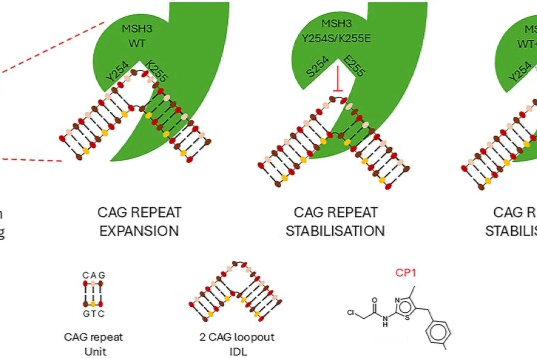
Genetic or pharmacological disruption of the MSH3 Y245/K246 IDL binding pocket slows CAG repeat expansion

Health economic evaluations of genomic newborn screening: Approaches by studies within the international consortium on newborn sequencing
Targeting toxic HTT1a could be the key to treating Huntington's disease

Spatial transcriptomics implicates the thalamus and cortex in autism and schizophrenia
The phenotypic spectrum and genetic determinants of severe spinal muscular atrophy in individuals with a single SMN2 copy: an international retrospective observational study
Personalized CRISPR therapies could soon reach thousands — here’s how