Centripetal Retinal Degeneration Follows an Exponential Decay and May Be Reversed Following Gene Therapy
Publication details
Summary
This is a study published in the American Journal of Ophthalmology. The study looked at people with a type of inherited retina disease called rod-cone dystrophy. The authors measured a bright ring seen on eye scans and found that it usually gets smaller in a very steady way over time. They also found that after gene therapy, this ring may get bigger again, which suggests the treatment may help the retina work better.
Keywords: rod-cone dystrophy, retinal degeneration, gene therapy, eye scans, retina, disease progression.
Abstract
Purpose
To model the progression of the hyperautofluorescent ring in rod-cone dystrophies and to report a novel finding of its reversal following gene therapy.
Design
Retrospective analysis of a prospective cohort study.
Participants
Fifty-eight individuals with genotyped rod-cone dystrophies and hyperautofluorescent rings on fundus autofluorescence (FAF).
Methods
Patients with a rod-cone dystrophy and hyperautofluorescent rings on FAF were identified and their rings categorized according to disease progression. Correlation between different FAF and optical coherence tomography (OCT) parameters in patients with early-mid stage disease were calculated. A retrospective, longitudinal analysis of FAF images was subsequently performed. A linear mixed model, with the dependent variable of total horizontal hyperautofluorescent ring width and independent variables of time, genotype and the interactions was used.
Main Outcome Measures
Horizontal diameter of the hyperautofluorescent ring.
Results
A total of 58 rod-cone dystrophy patients were analyzed in this study. Twenty eyes of 20 patients presented with early to mid-stage disease. The internal ring diameter, external ring diameter and ring thickness, were strongly correlated with the ellipsoid zone width (r = 0.94), external limiting membrane width (r = 0.97) and loss of the ellipsoid zone (r = 0.92) respectively. Longitudinal analysis suggests that FAF changes decline very predictably in a logarithmic fashion (R2 = 0.958). Results indicated a significant reversal of the hyperautofluorescent ring following RPGR gene therapy (using Cotoretigene Toliparvovec), correlating with improvements in retinal sensitivity.
Conclusions
Our findings suggest that centripetal retinal degeneration in rod-cone dystrophies follows an exponential decay and that gene therapy may reverse both structural and functional deficits.
Related publications
Perivascular Astrocytic Gliosis Induced by Retinal Gene Therapy
Beyond innovation: why rare disease gene therapy needs a reset
EQ-5D vision bolt-on in macular degeneration: associations with visual measures and effect on utility differences and cost-effectiveness of stereotactic radiotherapy
Clinical Spectrum and Genetics of Ocular Manifestations in Muscle Eye Brain Disease: A Literature Review
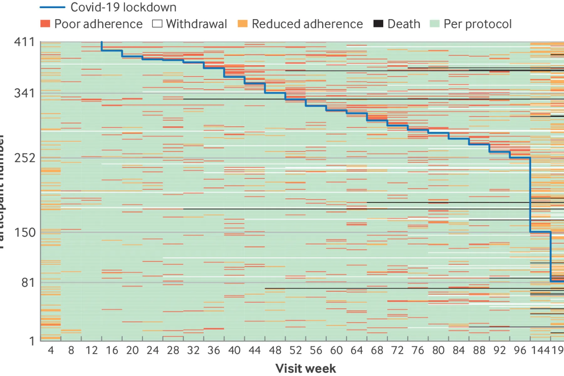
Stereotactic radiotherapy for neovascular age related macular degeneration: year 3 and 4 extended follow up results of a randomised, double masked, sham controlled, device trial (STAR)

Genomic newborn screening for actionable, sight-threatening eye diseases
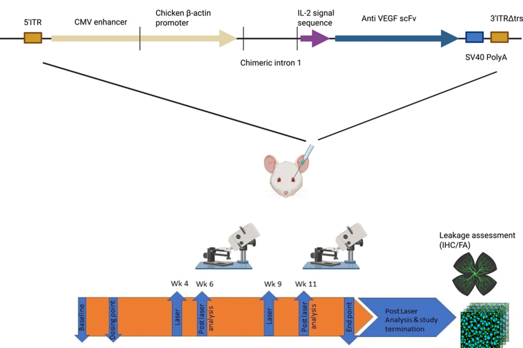
Efficacy of anti-VEGF single-chain variable fragment AAV-based gene therapy in a laser-induced choroidal neovascularisation mouse model

Health economic evaluations of genomic newborn screening: Approaches by studies within the international consortium on newborn sequencing
Personalized CRISPR therapies could soon reach thousands — here’s how