Perivascular Astrocytic Gliosis Induced by Retinal Gene Therapy
Publication details
Summary
Choroideremia is a rare inherited eye condition that causes people to slowly lose their sight. One possible treatment is gene therapy, where a harmless virus is used to carry a working gene into cells in the eye.
This study looked at 25 people who had received gene therapy for choroideremia. The researchers noticed a change in the retina, called perivascular astrocytic gliosis, in 12 people. This means that some support cells around the blood vessels in the retina became more crowded.
The change was seen only in people who received the higher dose of the gene therapy. It usually appeared within six months and then stayed stable. Importantly, the people affected did not have symptoms from it, and the researchers found no clear signs that it was caused by inflammation. The change also did not appear to get worse over time.
The researchers think this may be a harmless response to the gene therapy, but more research is needed to understand exactly why it happens.
Keywords
Choroideremia, retinal gene therapy, gene therapy, retina, AAV2, REP1, astrocytes, gliosis, vision, eye disease, retinal imaging, gene therapy safety
Abstract
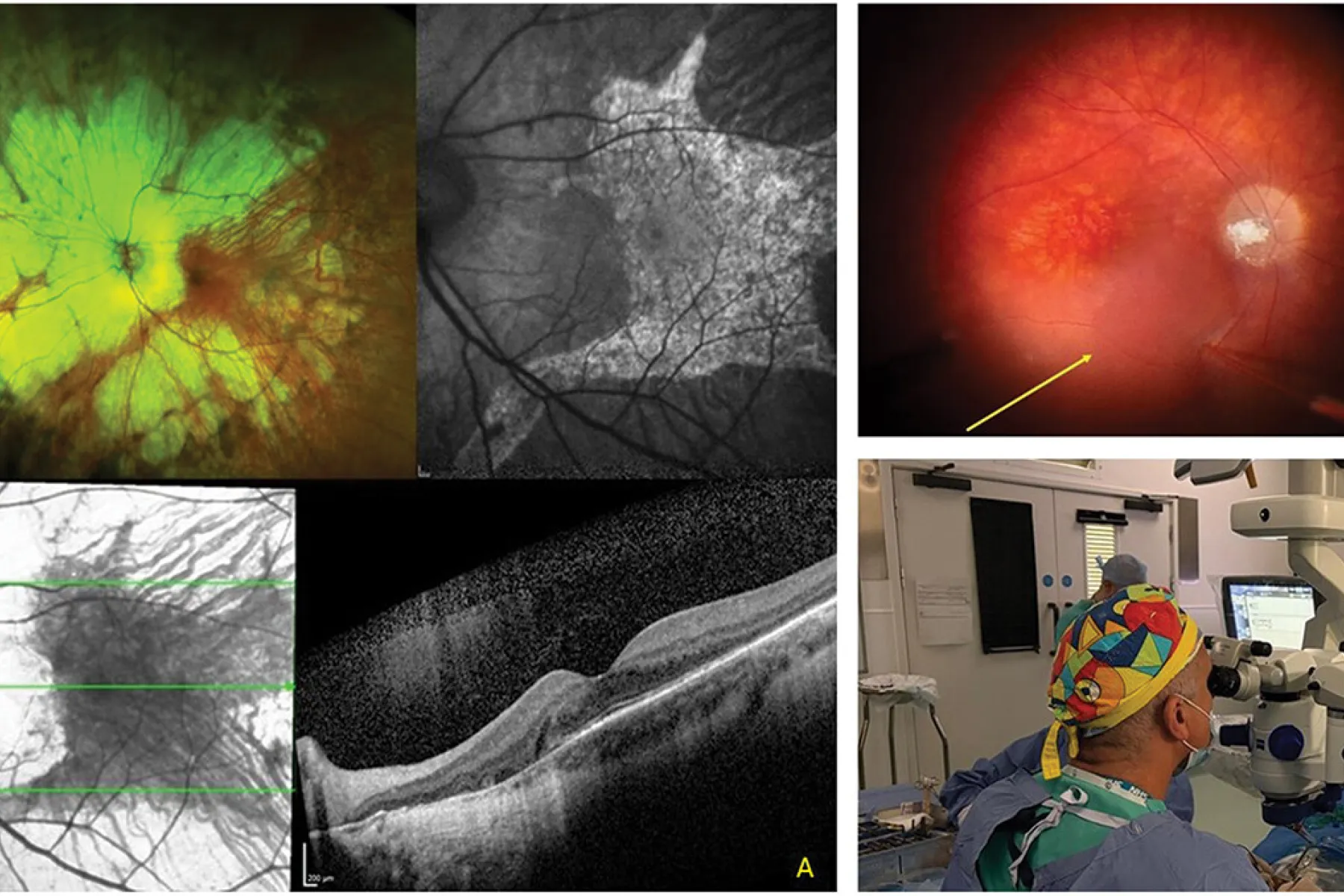
This retrospective follow-on study reviewed 25 consecutive patients with genetically confirmed choroideremia who were treated with subretinal AAV2-CAG-REP1 in dose-escalation gene therapy clinical trials conducted at Oxford University Hospitals NHS Foundation Trust and the University Eye Hospital Tübingen. Six patients in Oxford received 1010 vector genomes in 100 µL, while a further 19 patients across both centers received 1011 vector genomes in 100 µL. Although the primary safety outcomes of both investigator-led academic studies have previously been reported, retinal imaging changes were observed during follow-up, prompting a more detailed characterization of these findings. Twelve of 25 treated eyes developed imaging features consistent with perivascular astrocytic gliosis during follow-up, representing 48% of the cohort. All affected eyes had received the higher dose, accounting for 12 of 19 eyes treated with 1011 vector genomes. No patients receiving the lower dose (1.0 × 10¹⁰ vector genomes) developed this phenotype. The lesions followed the major vascular arcades toward the optic disc, and optical coherence tomography demonstrated inner retinal thickening with a fine, granular hyperreflective texture, without intraretinal cysts. Longitudinal imaging showed no progressive expansion of the involved regions. Clinically, the findings were silent: no patient reported uveitic symptoms, ocular examinations were largely quiet, and no peripheral cellular immune responses were detected in patients with available assays. Perivascular astrocytic gliosis therefore appears to be a common, clinically silent, nonprogressive, and noninflammatory retinal response after subretinal AAV2-CAG-REP1 gene therapy for choroideremia at a dose of 1011 vector genomes, which is generally accepted as the therapeutic human dose for targeting photoreceptors. We hypothesize that local REP1 overexpression in perivascular astrocytes suppresses FOXO3-mediated apoptosis and engages EGFR/STAT3 signaling, driving benign perivascular astrocytic accumulation analogous to retinal astrocytic hamartomas seen in phacomatoses. Gene therapy involving ubiquitous promoters may have unexpected effects, and while benign, may not be predicted.
Related publications
Beyond innovation: why rare disease gene therapy needs a reset
EQ-5D vision bolt-on in macular degeneration: associations with visual measures and effect on utility differences and cost-effectiveness of stereotactic radiotherapy
Clinical Spectrum and Genetics of Ocular Manifestations in Muscle Eye Brain Disease: A Literature Review
Stereotactic radiotherapy for neovascular age related macular degeneration: year 3 and 4 extended follow up results of a randomised, double masked, sham controlled, device trial (STAR)

Genomic newborn screening for actionable, sight-threatening eye diseases
Efficacy of anti-VEGF single-chain variable fragment AAV-based gene therapy in a laser-induced choroidal neovascularisation mouse model

Health economic evaluations of genomic newborn screening: Approaches by studies within the international consortium on newborn sequencing
Centripetal Retinal Degeneration Follows an Exponential Decay and May Be Reversed Following Gene Therapy
Personalized CRISPR therapies could soon reach thousands — here’s how